
Definition of endometrial cancer: Cancer that forms in the tissue lining the uterus (the small, hollow, pear-shaped organ in a woman's pelvis in which a fetus develops). Most endometrial cancers are adenocarcinomas (cancers that begin in cells that make and release mucus and other fluids).
Endometrial cancer is a disease in which malignant (cancer) cells form in the tissues of the endometrium.
The endometrium is the lining of the uterus, a hollow, muscular organ in a woman’s pelvis. The uterus is where a fetus grows. In most nonpregnant women, the uterus is about 3 inches long. The lower, narrow end of the uterus is the cervix, which leads to the vagina.

Anatomy of the female reproductive system. The organs in the female reproductive system include the uterus, ovaries, fallopian tubes, cervix, and vagina. The uterus has a muscular outer layer called the myometrium and an inner lining called the endometrium.
Taking tamoxifen for breast cancer or taking estrogen alone (without progesterone) can increase the risk of endometrial cancer.
Endometrial cancer may develop in breast cancer patients who have been treated with tamoxifen. A patient taking this drug should have a pelvic exam every year and report any vaginal bleeding (other than menstrual bleeding) as soon as possible. Women taking estrogen (a hormone that can affect the growth of some cancers) alone have an increased risk of endometrial cancer. Taking estrogen combined with progesterone (another hormone) does not increase a woman’s risk of this cancer.
These and other symptoms may be caused by endometrial cancer. Other conditions may cause the same symptoms. Check with your doctor if you have any of the following problems:
- Bleeding or discharge not related to menstruation (periods).
- Difficult or painful urination.
- Pain during sexual intercourse.
- Pain in the pelvic area.
Because endometrial cancer begins inside the uterus, it does not usually show up in the results of aPap test. For this reason, a sample of endometrial tissue must be removed and checked under amicroscope to look for cancer cells. One of the following procedures may be used:
- Endometrial biopsy : The removal of tissue from the endometrium (inner lining of the uterus) by inserting a thin, flexible tube through the cervix and into the uterus. The tube is used to gently scrape a small amount of tissue from the endometrium and then remove the tissue samples. Apathologist views the tissue under a microscope to look for cancer cells.
- Dilatation and curettage : A procedure to remove samples of tissue from the inner lining of the uterus. The cervix is dilated and a curette (spoon-shaped instrument) is inserted into the uterus to remove tissue. The tissue samples are checked under a microscope for signs of disease. This procedure is also called a D&C.
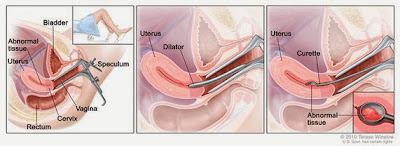
- Dilatation and curettage (D and C). A speculum is inserted into the vagina to widen it in order to look at the cervix (first panel). A dilator is used to widen the cervix (middle panel). A curette is put through the cervix into the uterus to scrape out abnormal tissue (last panel).

No comments:
Post a Comment